No products in the cart.
TECHNOLOGY
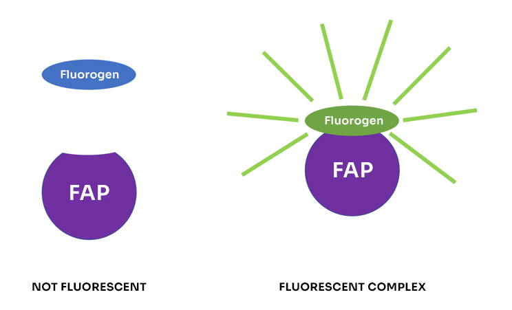
Fluorogen-activating proteins – FAPs – are polypeptides that bind organic fluorogen molecules at nanomolar concentrations to yield fluorescent complexes. Neither the FAP nor the fluorogen is fluorescent by itself.

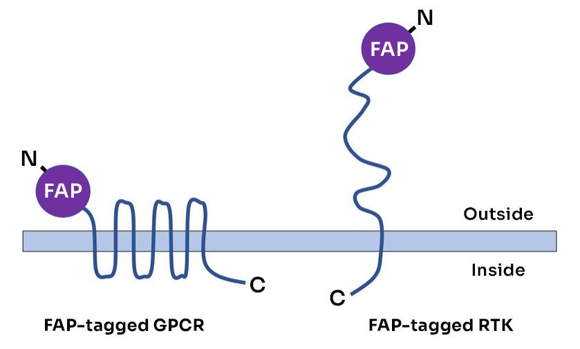
Agonist stimulation of G-protein coupled receptors (GPCRs) or receptor tyrosine kinases (RTKs) typically triggers receptor internalization. FAP-based assays are particularly useful for detecting and quantifying this response. For these assays, receptors are expressed with FAP tags at their extracellular N-termini.

When cells expressing FAP-tagged receptors are exposed to membrane impermeant fluorogens, receptor molecules at the cell surface acquire fluorescence but receptor molecules inside the cell do not. If fluorescence subsequently appears inside the cell, it must come from receptor molecules that were internalized after fluorogen addition.
To detect and quantify receptor internalization, we developed two new fluorogens for which fluorescent signal originating from receptors at the cell surface differs in spectral properties from signal originating from internalized receptors. These proprietary fluorogens enable the live-cell assays described below.
The pHD Assay
The pHD assay uses a proprietary fluorogen (αRED-pHD) that gives direct red fluorescence (ex=640nm/ em=680nm) and indirect (FRET) fluorescence (ex=560nm/ em=680nm). The FRET signal – but not the red signal – is enhanced at acidic pH, and so receptor molecules that have been internalized in acidified endosomes show higher FRET than receptor molecules at the surface. This enables one to quantify the fraction of receptors that have been internalized by measuring the FRET-to-RED ratio for the cell as a whole.
In brief, the assay is performed as follows:
Step 1. Add 100nM pHD fluorogen to cells expressing alphaFAP-tagged receptor
Step 2. Add test compound
Step 3. Incubate at 37o
Step 4. Measure signal in RED and FRET channels to detect and quantify receptor internalization
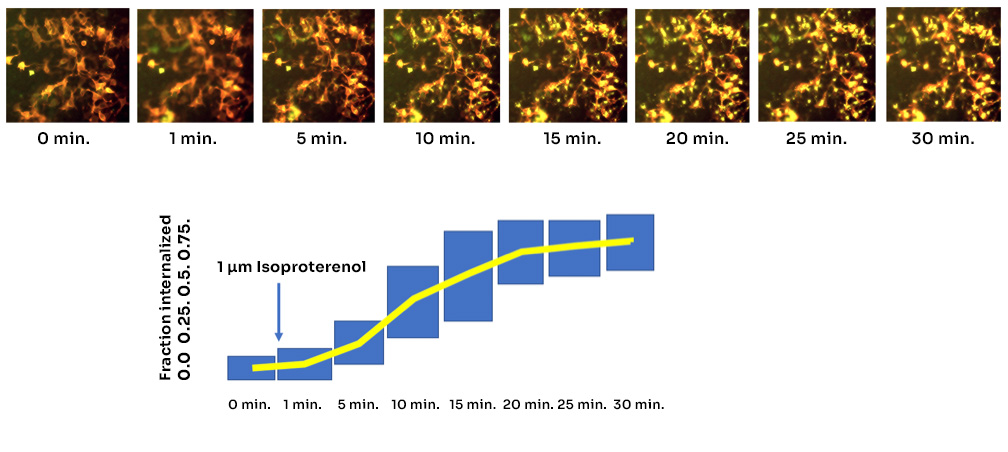
The pHD assay is a real-time assay that gives kinetic output. An example is shown below for isoproterenol stimulation of cells that express a FAP-tagged human adrenergic receptor (ADRB2). Direct signal is displayed in red in the images, and FRET signal is displayed in green. The box plots in the figure represent the median 75% of all cells at each time point.

The SSC Assay
The SSC assay uses a proprietary fluorogen (αRED-SSC) that, like the pHD fluorogen, gives direct fluorescence and indirect FRET fluorescence, both at 680nm. The SSC fluorogen carries a disulfide bond that can be cleaved by a reducing agent to release the FRET donor moiety and eliminate FRET signal. Treatment of cells with a membrane-impermeant reducing agent eliminates FRET signal from receptors at the cell surface but does not affect FRET signal from internalized receptors.
In brief, the assay is performed as follows:
Step 1. Add 100nM SSC fluorogen to cultured cells expressing alphaFAP-tagged receptor
Step 2. Add test compound
Step 3. Incubate for 10-40 minutes
Step 4. Add reducing agent
Step 5. Add fixative
Step 6. Measure signal in RED and FRET channels to detect and quantify receptor internalization.
Because cells are fixed before measurement, the SSC assay allows one to separate the wet-lab steps from the analytical steps of the assay. Example results are shown below for HEK 293 cells expressing the human gastric inhibitory polypeptide receptor (GIPR). Compared to controls, cells treated with agonist (5 micromolar GIP for 40 minutes) show a nearly twofold increase in the FRET-to-RED ratio (0.75 versus 0.38) indicating that nearly half of the surface receptors were internalized.

| Red Signal | Background | Red Minus BKD | FRET Signal | Background | FRET Minus BKD | FRET Minus BKD/Red Minus BKD | |
|---|---|---|---|---|---|---|---|
| No drug, 40′ | 14072 | 10684 | 3388 | 11810 | 10532 | 1278 | 0.38 |
| 5 µM GIP, 40′ | 14876 | 11404 | 3472 | 15149 | 12549 | 2600 | 0.75 |
Please contact us at info@spectragenetics.com to request additional information about these assays. We will be pleased to hear from you and to help with your assay needs.
SpectraGenetics supplies two classes of FAP – alpha and beta – and a variety of fluorogens specific to each. Key properties of these molecules are summarized in the tables below.
Our products include mammalian cell lines that express FAP-tagged receptors [link], expression vectors for FAP-tagged receptors [link], and a variety of fluorogen molecules [link]. If you are interested in a receptor that is not in our catalog, we can make a cell line or expression vector for you on a custom basis [link].
| FAP | Kd for Fluorogens |
|---|---|
| Alpha1 | 3.2 nM for αRED-np and αRED-p |
| Alpha2 | <1 nM for αRED; 3 nM for αRED-pH and αRED-SS |
| Beta1 | 3.1 nM for βGREEN; 13.5 nM for βRED |
| Beta2 | 360 nM for βGREEN |
| FLUOROGEN | Ex-Max | Em-Max |
|---|---|---|
| αRED-np | 640 nm | 680 nm |
| αRED-p | 640 nm | 680 nm |
| αRED-pHD | 560 and 640 nm | 680 nm |
| αRED-SSC | 560 and 640 nm | 680 nm |
| βGREEN-np | 510 nm | 530 nm |
| βRED-np | 510 and 650 nm | 680 nm |
The seminal paper describing the FAPs and fluorogens can be seen here, and a review describing a range of FAP applications is here.
A bibliography of nearly 100 papers describing FAP-based assays and other FAP applications can be viewed here. In these papers the FAPs and fluorogens are denoted by a number of different names. The table below relates those names to the names that we use in our products.
| SpectraGenetics Product Name | Alternative Name |
|---|---|
| FAP alpha1 | MG13, HL4-MG |
| FAP alpha2 | L5-dimer, dL5, dNP138 |
| FAP beta1 | HL1.0.1-TO1, AMII.2 |
| FAP beta2 | HL1-TO1, scFv1 |
| αRED-np | MG-Btau |
| αRED-p | MG-ester |
| βGREEN-np | TO-2p |
| βRED-np | TO-2p-Cy5 |
FAP tags were developed at Carnegie Mellon University and are the subjects of multiple patents including US8664364, US9023998, US9249306, US9688743, US9995679, and foreign equivalents. SpectraGenetics is the exclusive world-wide commercial licensee of these patents in the field of GPCR assays.


